- Overview
- Log In For Videos
- Give Feedback
- Seizure Classification
- Unknown Onset Seizure
- Neonatal Seizure
- Epilepsy Classification
- Generalized Epilepsy
- Focal Epilepsy
- Generalized and Focal Epilepsy
- Unknown Epilepsy
- Epilepsy Syndromes
- Epilepsy Etiologies
- Metabolic Etiologies
- Immune Etiologies
- Infectious Etiologies
- Unknown Etiologies
- Encephalopathy
- Epilepsy imitators
SUBCORTICAL BAND HETEROTOPIA
Imaging
Imaging for optimized detection of subcortical band heterotopia:
Whilst subcortical band heterotopia may be seen on USS and CT (depending on severity), MRI is the imaging of choice for assessing the detail and associated structural abnormalities. MRI should include thin slice volumetric T1-weighted images, axial and coronal T2-weighted and FLAIR images.
Imaging characteristics of subcortical band heterotopia:
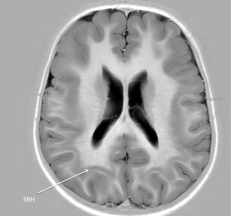
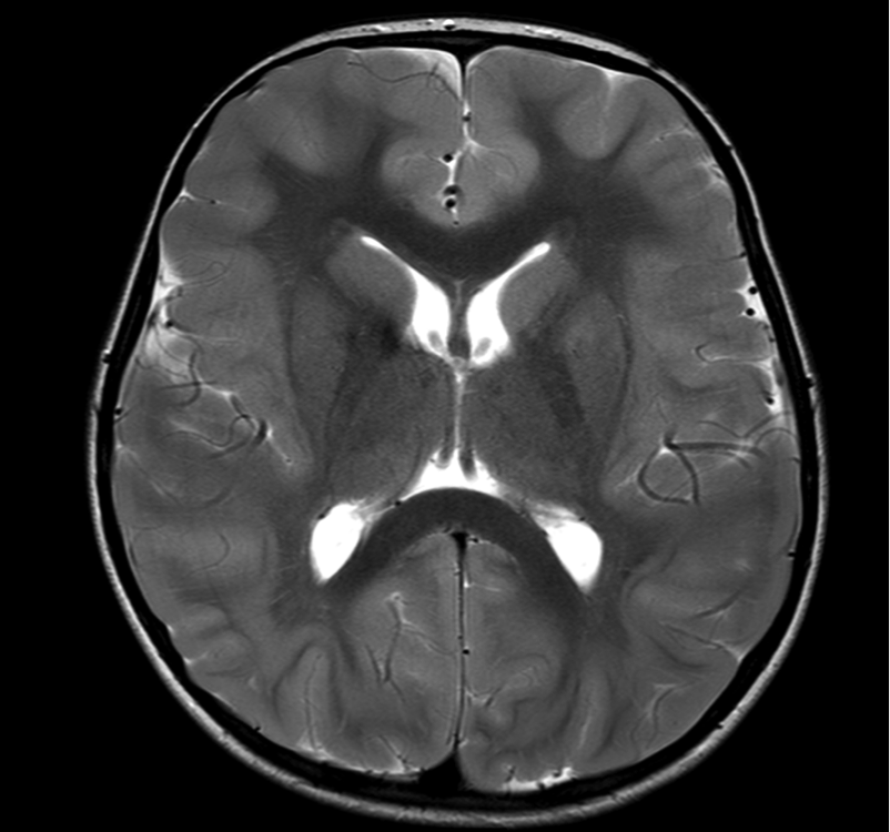
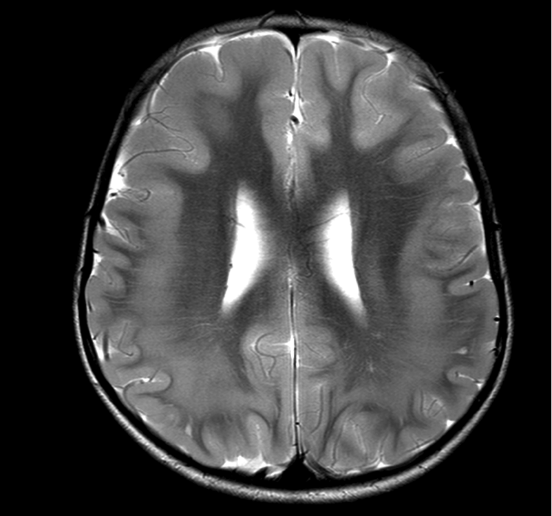
- a band of grey matter is seen, located between the ventricle and cortex, this can be of variable thickness (can be subtle in some areas), and may be duplicated (i.e. a double band) in the temporal lobes
- the overlying cortex may be normal or may show shallow sulcation.
- Anterior predominance of banding is recognized with DCX pathogenic variants, posterior predominance with LIS1 pathogenic variants
Imaging in subcortical band heterotopia
The images below are all from the same patient, and show pachygyria (broad simple gyri) anteriorly and subcortical band heterotopia posteriorly (SBH, arrows).
Feedback
|
Home
|
Contact Us
|
Privacy
|
Terms & Conditions of Use
|
Log In For Videos

This website is owned by the International League Against Epilepsy. Text on this website, last updated June 30, 2024,
is available under a Creative Commons Attribution-ShareAlike 4.0 International License,
EXCEPTING all videos and images, which remain copyrighted by the International League Against Epilepsy.