- Overview
- Log In For Videos
- Give Feedback
- Seizure Classification
- Unknown Onset Seizure
- Neonatal Seizure
- Epilepsy Classification
- Generalized Epilepsy
- Focal Epilepsy
- Generalized and Focal Epilepsy
- Unknown Epilepsy
- Epilepsy Syndromes
- Epilepsy Etiologies
- Metabolic Etiologies
- Immune Etiologies
- Infectious Etiologies
- Unknown Etiologies
- Encephalopathy
- Epilepsy imitators
GREY MATTER HETEROTOPIA
Imaging
Imaging for optimized detection of grey matter heterotopia:
Whilst grey matter heterotopia may be seen on USS and CT (depending on size), MRI is the imaging of choice for assessing the detail and associated structural abnormalities. MRI should include thin slice volumetric T1-weighted images, axial and coronal T2-weighted and FLAIR images.
Imaging characteristics of periventricular nodular heterotopia:
- heterotopic nodules of grey matter intensity are seen
immediately deep to the ependymal layer, two patterns are seen:
- nodules along the bodies and anterior horns of the lateral ventricles, as seen in typical bilateral periventricular nodular heterotopia
- nodules seen maximal in the trigones and occipital horns, as seen in the infrasylvian form of periventricular nodular heterotopia
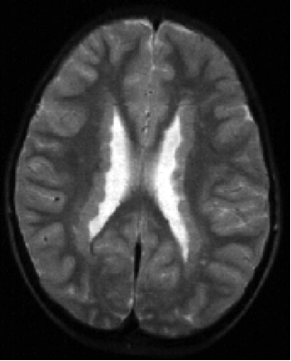
Imaging in grey matter heterotopia
The image below is an example of bilateral periventricular nodular heterotopia, showing grey matter nodules along the bodies of both lateral ventricles.
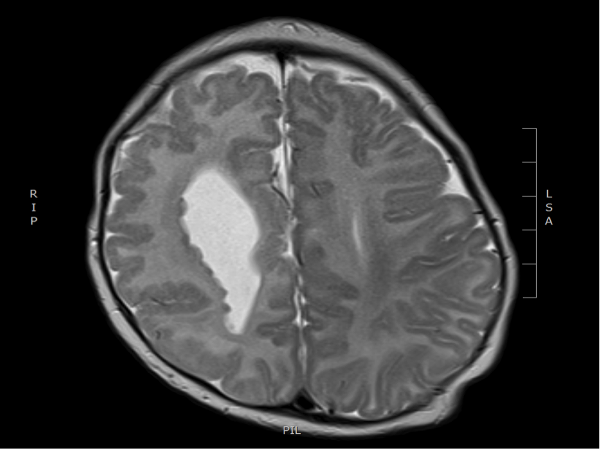
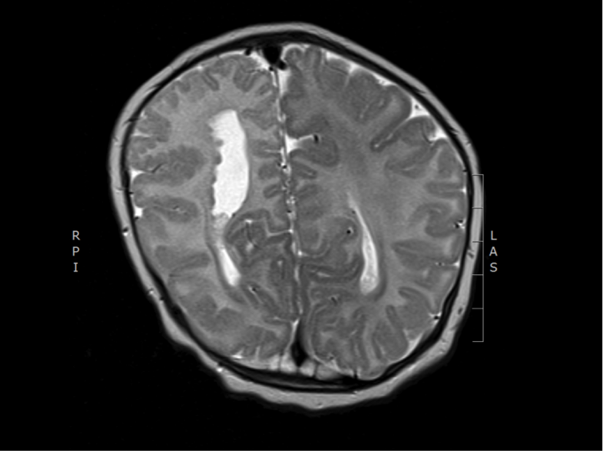
Imaging in grey matter heterotopia
Both images below are from the same patient, and show unilateral (right) periventricular nodular heterotopia, with grey matter heterotopia lining the body of the right ventricle. There is polymicrogyria in the overlying cortex (compare right and left).
Imaging characteristics of subcortical nodular heterotopia:
- heterotopic grey matter extending from the ventricle into white matter or extending from the cortex into underlying white matter
- heterotopias are continuous with overlying cortex or underlying ventricle
- co-occurring abnormalities in the affected hemisphere include distortion of the ventricle, loss of volume in cortex and white matter, abnormal white matter signal
Feedback
|
Home
|
Contact Us
|
Privacy
|
Terms & Conditions of Use
|
Log In For Videos

This website is owned by the International League Against Epilepsy. Text on this website, last updated June 30, 2024,
is available under a Creative Commons Attribution-ShareAlike 4.0 International License,
EXCEPTING all videos and images, which remain copyrighted by the International League Against Epilepsy.