
- Overview
- Log In For Videos
- Give Feedback
- Seizure Classification
- Unknown Onset Seizure
- Neonatal Seizure
- Epilepsy Classification
- Generalized Epilepsy
- Focal Epilepsy
- Generalized and Focal Epilepsy
- Unknown Epilepsy
- Epilepsy Syndromes
- Epilepsy Etiologies
- Metabolic Etiologies
- Immune Etiologies
- Infectious Etiologies
- Unknown Etiologies
- Encephalopathy
- Epilepsy imitators
RASMUSSEN SYNDROME (RS)
Imaging
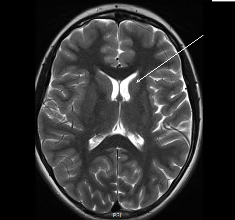
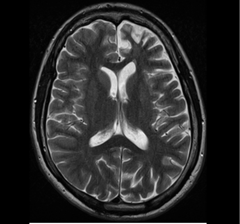
Progressive atrophy of one hemisphere on serial neuroimaging is the hallmark of Rasmussen syndrome. This may not be evident in the initial prodromal phase of the illness, however within months of onset of the acute stage, most patients show unilateral enlargement of the outer and inner CSF compartments. The loss of cerebral volume often starts in the insular region, with enlargement of the temporal horn of the ventricle and the Sylvian fissure. Ipsilateral atrophy of the head of the caudate nucleus is another early diagnostic clue. T2/FLAIR hyperintense signal abnormality is seen in cortical or subcortical regions, or both. The occurrence of bilateral disease is debated but is certainly very rare.
Imaging of Rasmussen syndrome
Both images below are from the same patient and illustrate the findings of Rasmussen syndrome over time. The first image shows mild reduction in volume of the left hemisphere, with atrophy of the head of caudate (arrow). The second image shows progression of the atrophy over time, with atrophy of the head of caudate also seen on this image.

